Search Results for author:
Found 14 papers, 2 papers with code
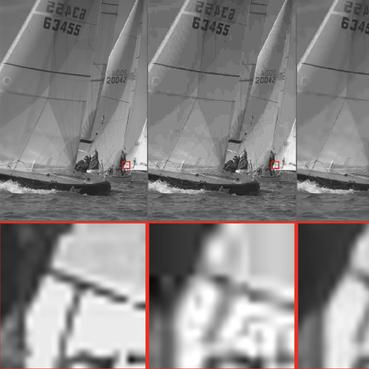
Micro CT Image-Assisted Cross Modality Super-Resolution of Clinical CT Images Utilizing Synthesized Training Dataset
Unsupervised SR methods are required that do not need paired LR and HR images.

Super-resolution of clinical CT volumes with modified CycleGAN using micro CT volumes
This paper presents a super-resolution (SR) method with unpaired training dataset of clinical CT and micro CT volumes.
Visualizing intestines for diagnostic assistance of ileus based on intestinal region segmentation from 3D CT images
This paper presents a visualization method of intestine (the small and large intestines) regions and their stenosed parts caused by ileus from CT volumes.
Multi-modality super-resolution loss for GAN-based super-resolution of clinical CT images using micro CT image database
This paper newly introduces multi-modality loss function for GAN-based super-resolution that can maintain image structure and intensity on unpaired training dataset of clinical CT and micro CT volumes.

A multi-scale pyramid of 3D fully convolutional networks for abdominal multi-organ segmentation
Recent advances in deep learning, like 3D fully convolutional networks (FCNs), have improved the state-of-the-art in dense semantic segmentation of medical images.
Unsupervised Segmentation of 3D Medical Images Based on Clustering and Deep Representation Learning
This paper presents a novel unsupervised segmentation method for 3D medical images.

Unsupervised Pathology Image Segmentation Using Representation Learning with Spherical K-means
In this paper, we propose a unified approach to unsupervised representation learning and clustering for pathology image segmentation.
Deep learning and its application to medical image segmentation
However, recent advances in deep learning have made it possible to significantly improve the performance of image recognition and semantic segmentation methods in the field of computer vision.
An application of cascaded 3D fully convolutional networks for medical image segmentation
In this work, we show that a multi-class 3D FCN trained on manually labeled CT scans of several anatomical structures (ranging from the large organs to thin vessels) can achieve competitive segmentation results, while avoiding the need for handcrafting features or training class-specific models.
 Ranked #2 on
3D Medical Imaging Segmentation
on TCIA Pancreas-CT
Ranked #2 on
3D Medical Imaging Segmentation
on TCIA Pancreas-CT

On the influence of Dice loss function in multi-class organ segmentation of abdominal CT using 3D fully convolutional networks
Deep learning-based methods achieved impressive results for the segmentation of medical images.
Towards dense volumetric pancreas segmentation in CT using 3D fully convolutional networks
Pancreas segmentation in computed tomography imaging has been historically difficult for automated methods because of the large shape and size variations between patients.

Towards Automatic Abdominal Multi-Organ Segmentation in Dual Energy CT using Cascaded 3D Fully Convolutional Network
In this paper, we proposed a 3D FCN based method for automatic multi-organ segmentation in DECT.
Hierarchical 3D fully convolutional networks for multi-organ segmentation
In this work, we show that a multi-class 3D FCN trained on manually labeled CT scans of seven abdominal structures (artery, vein, liver, spleen, stomach, gallbladder, and pancreas) can achieve competitive segmentation results, while avoiding the need for handcrafting features or training organ-specific models.
Multi-scale Image Fusion Between Pre-operative Clinical CT and X-ray Microtomography of Lung Pathology
The alignment of clinical CT with $\mu$CT will allow further registration with even finer resolutions of $\mu$CT (up to 10 $\mu$m resolution) and ultimately with histopathological microscopy images for further macro to micro image fusion that can aid medical image analysis.
