Search Results for author:
Found 24 papers, 4 papers with code
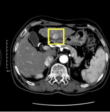
A Flexible Three-Dimensional Hetero-phase Computed Tomography Hepatocellular Carcinoma (HCC) Detection Algorithm for Generalizable and Practical HCC Screening
We develop a flexible three-dimensional deep algorithm, called hetero-phase volumetric detection (HPVD), that can accept any combination of contrast-phase inputs and with adjustable sensitivity depending on the clinical purpose.


Lesion Segmentation and RECIST Diameter Prediction via Click-driven Attention and Dual-path Connection
PDNet learns comprehensive and representative deep image features for our tasks and produces more accurate results on both lesion segmentation and RECIST diameter prediction.

Weakly-Supervised Universal Lesion Segmentation with Regional Level Set Loss
Accurately segmenting a variety of clinically significant lesions from whole body computed tomography (CT) scans is a critical task on precision oncology imaging, denoted as universal lesion segmentation (ULS).
Fully-Automated Liver Tumor Localization and Characterization from Multi-Phase MR Volumes Using Key-Slice ROI Parsing: A Physician-Inspired Approach
Using radiological scans to identify liver tumors is crucial for proper patient treatment.
Deep Lesion Tracker: Monitoring Lesions in 4D Longitudinal Imaging Studies
In this work, we present deep lesion tracker (DLT), a deep learning approach that uses both appearance- and anatomical-based signals.

SAM: Self-supervised Learning of Pixel-wise Anatomical Embeddings in Radiological Images
We introduce such an approach, called Self-supervised Anatomical eMbedding (SAM).
User-Guided Domain Adaptation for Rapid Annotation from User Interactions: A Study on Pathological Liver Segmentation
Mask-based annotation of medical images, especially for 3D data, is a bottleneck in developing reliable machine learning models.

Learning from Multiple Datasets with Heterogeneous and Partial Labels for Universal Lesion Detection in CT
For example, DeepLesion is such a large-scale CT image dataset with lesions of various types, but it also has many unlabeled lesions (missing annotations).
Deep Volumetric Universal Lesion Detection using Light-Weight Pseudo 3D Convolution and Surface Point Regression
Identifying, measuring and reporting lesions accurately and comprehensively from patient CT scans are important yet time-consuming procedures for physicians.
Lymph Node Gross Tumor Volume Detection in Oncology Imaging via Relationship Learning Using Graph Neural Network
Specifically, we first utilize a 3D convolutional neural network with ROI-pooling to extract the GTV$_{LN}$'s instance-wise appearance features.
Harvesting, Detecting, and Characterizing Liver Lesions from Large-scale Multi-phase CT Data via Deep Dynamic Texture Learning
To this end, we propose a fully-automated and multi-stage liver tumor characterization framework designed for dynamic contrast computed tomography (CT).
Uncertainty-aware multi-view co-training for semi-supervised medical image segmentation and domain adaptation
Experiments on the NIH pancreas segmentation dataset and a multi-organ segmentation dataset show state-of-the-art performance of the proposed framework on semi-supervised medical image segmentation.
Universal Lesion Detection by Learning from Multiple Heterogeneously Labeled Datasets
First, we learn a multi-head multi-task lesion detector using all datasets and generate lesion proposals on DeepLesion.
 Ranked #5 on
Medical Object Detection
on DeepLesion
(using extra training data)
Ranked #5 on
Medical Object Detection
on DeepLesion
(using extra training data)
Co-Heterogeneous and Adaptive Segmentation from Multi-Source and Multi-Phase CT Imaging Data: A Study on Pathological Liver and Lesion Segmentation
In medical imaging, organ/pathology segmentation models trained on current publicly available and fully-annotated datasets usually do not well-represent the heterogeneous modalities, phases, pathologies, and clinical scenarios encountered in real environments.
Detecting Scatteredly-Distributed, Small, andCritically Important Objects in 3D OncologyImaging via Decision Stratification
We focus on the detection and segmentation of oncology-significant (or suspicious cancer metastasized) lymph nodes (OSLNs), which has not been studied before as a computational task.
Lesion Harvester: Iteratively Mining Unlabeled Lesions and Hard-Negative Examples at Scale
This is the goal of our work, where we develop a powerful system to harvest missing lesions from the DeepLesion dataset at high precision.
End-to-End Adversarial Shape Learning for Abdomen Organ Deep Segmentation
However, it is challenging to train the conventional CNN-based segmentation models that aware of the shape and topology of organs.

3D Semi-Supervised Learning with Uncertainty-Aware Multi-View Co-Training
Meanwhile, a fully-supervised method based on our approach achieved state-of-the-art performances on both the LiTS liver tumor segmentation and the Medical Segmentation Decathlon (MSD) challenge, demonstrating the robustness and value of our framework, even when fully supervised training is feasible.

CT Image Enhancement Using Stacked Generative Adversarial Networks and Transfer Learning for Lesion Segmentation Improvement
The first GAN reduces the noise in the CT image and the second GAN generates a higher resolution image with enhanced boundaries and high contrast.
Iterative Attention Mining for Weakly Supervised Thoracic Disease Pattern Localization in Chest X-Rays
Given image labels as the only supervisory signal, we focus on harvesting, or mining, thoracic disease localizations from chest X-ray images.


Accurate Weakly-Supervised Deep Lesion Segmentation using Large-Scale Clinical Annotations: Slice-Propagated 3D Mask Generation from 2D RECIST
Volumetric lesion segmentation from computed tomography (CT) images is a powerful means to precisely assess multiple time-point lesion/tumor changes.
Pancreas Segmentation in CT and MRI Images via Domain Specific Network Designing and Recurrent Neural Contextual Learning
Automatic pancreas segmentation in radiology images, eg., computed tomography (CT) and magnetic resonance imaging (MRI), is frequently required by computer-aided screening, diagnosis, and quantitative assessment.
Accurate Weakly Supervised Deep Lesion Segmentation on CT Scans: Self-Paced 3D Mask Generation from RECIST
Toward this end, we introduce a convolutional neural network based weakly supervised self-paced segmentation (WSSS) method to 1) generate the initial lesion segmentation on the axial RECIST-slice; 2) learn the data distribution on RECIST-slices; 3) adapt to segment the whole volume slice by slice to finally obtain a volumetric segmentation.
Improving Deep Pancreas Segmentation in CT and MRI Images via Recurrent Neural Contextual Learning and Direct Loss Function
The output layer of this network module is then connected to recurrent layers and can be fine-tuned for contextual learning, in an end-to-end manner.
