Search Results for author:
Found 33 papers, 4 papers with code
ConDistFL: Conditional Distillation for Federated Learning from Partially Annotated Data
Federated learning (FL) is a key technology enabling the collaborative development of a model without exchanging training data.

Identifying Suspicious Regions of Covid-19 by Abnormality-Sensitive Activation Mapping
This paper presents a fully-automated method for the identification of suspicious regions of a coronavirus disease (COVID-19) on chest CT volumes.
KST-Mixer: Kinematic Spatio-Temporal Data Mixer For Colon Shape Estimation
Kinematic data of a colonoscope and the colon, including positions and directions of their centerlines, are obtained using electromagnetic and depth sensors.
Semi-automated Virtual Unfolded View Generation Method of Stomach from CT Volumes
In this paper, we propose a semi-automated method for generating VU views of the stomach.
Realistic Endoscopic Image Generation Method Using Virtual-to-real Image-domain Translation
Virtual endoscopic images are generated by using a volume rendering method from a CT volume of a patient.

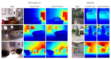
Depth Estimation from Single-shot Monocular Endoscope Image Using Image Domain Adaptation And Edge-Aware Depth Estimation
The identification accuracy of the network improved from 69. 2% to 74. 1% by using the estimated depth images.

COVID-19 Infection Segmentation from Chest CT Images Based on Scale Uncertainty
We utilize the scale uncertainty among various receptive field sizes of a segmentation FCN to obtain infection regions.
Lung infection and normal region segmentation from CT volumes of COVID-19 cases
Our method recognizes and segments lung normal and infection regions in CT volumes.
Multi-task Federated Learning for Heterogeneous Pancreas Segmentation
Federated learning (FL) for medical image segmentation becomes more challenging in multi-task settings where clients might have different categories of labels represented in their data.
Micro CT Image-Assisted Cross Modality Super-Resolution of Clinical CT Images Utilizing Synthesized Training Dataset
Unsupervised SR methods are required that do not need paired LR and HR images.
Automated Pancreas Segmentation Using Multi-institutional Collaborative Deep Learning
The performance of deep learning-based methods strongly relies on the number of datasets used for training.
Regression Forest-Based Atlas Localization and Direction Specific Atlas Generation for Pancreas Segmentation
This paper proposes a fully automated atlas-based pancreas segmentation method from CT volumes utilizing atlas localization by regression forest and atlas generation using blood vessel information.

Automated eye disease classification method from anterior eye image using anatomical structure focused image classification technique
This paper presents an automated classification method of infective and non-infective diseases from anterior eye images.


Colon Shape Estimation Method for Colonoscope Tracking using Recurrent Neural Networks
We propose a colon deformation estimation method using RNN and obtain the colonoscope shape from electromagnetic sensors during its insertion into the colon.
Colonoscope tracking method based on shape estimation network
We utilize the shape estimation network (SEN), which estimates deformed colon shape during colonoscope insertions.
Super-resolution of clinical CT volumes with modified CycleGAN using micro CT volumes
This paper presents a super-resolution (SR) method with unpaired training dataset of clinical CT and micro CT volumes.
Organ Segmentation From Full-size CT Images Using Memory-Efficient FCN
In this paper, we present a memory-efficient FCN to tackle the high GPU memory demand challenge in organ segmentation problem from clinical CT images.

Visualizing intestines for diagnostic assistance of ileus based on intestinal region segmentation from 3D CT images
This paper presents a visualization method of intestine (the small and large intestines) regions and their stenosed parts caused by ileus from CT volumes.
Multi-modality super-resolution loss for GAN-based super-resolution of clinical CT images using micro CT image database
This paper newly introduces multi-modality loss function for GAN-based super-resolution that can maintain image structure and intensity on unpaired training dataset of clinical CT and micro CT volumes.
Precise Estimation of Renal Vascular Dominant Regions Using Spatially Aware Fully Convolutional Networks, Tensor-Cut and Voronoi Diagrams
Then we generate a Voronoi diagram to estimate the renal vascular dominant regions based on the segmented kidney and renal arteries.
3D FCN Feature Driven Regression Forest-Based Pancreas Localization and Segmentation
We estimate the position and the size of the pancreas (localized) from global features by regression forests.
Machine learning-based colon deformation estimation method for colonoscope tracking
An estimation method of colon deformations occur during colonoscope insertions is necessary to reduce tracking errors.
A multi-scale pyramid of 3D fully convolutional networks for abdominal multi-organ segmentation
Recent advances in deep learning, like 3D fully convolutional networks (FCNs), have improved the state-of-the-art in dense semantic segmentation of medical images.
Unsupervised Pathology Image Segmentation Using Representation Learning with Spherical K-means
In this paper, we propose a unified approach to unsupervised representation learning and clustering for pathology image segmentation.

Unsupervised Segmentation of 3D Medical Images Based on Clustering and Deep Representation Learning
This paper presents a novel unsupervised segmentation method for 3D medical images.
Deep learning and its application to medical image segmentation
However, recent advances in deep learning have made it possible to significantly improve the performance of image recognition and semantic segmentation methods in the field of computer vision.
An application of cascaded 3D fully convolutional networks for medical image segmentation
In this work, we show that a multi-class 3D FCN trained on manually labeled CT scans of several anatomical structures (ranging from the large organs to thin vessels) can achieve competitive segmentation results, while avoiding the need for handcrafting features or training class-specific models.
 Ranked #2 on
3D Medical Imaging Segmentation
on TCIA Pancreas-CT
Ranked #2 on
3D Medical Imaging Segmentation
on TCIA Pancreas-CT

On the influence of Dice loss function in multi-class organ segmentation of abdominal CT using 3D fully convolutional networks
Deep learning-based methods achieved impressive results for the segmentation of medical images.
Towards dense volumetric pancreas segmentation in CT using 3D fully convolutional networks
Pancreas segmentation in computed tomography imaging has been historically difficult for automated methods because of the large shape and size variations between patients.
Airway segmentation from 3D chest CT volumes based on volume of interest using gradient vector flow
In this paper, we propose a new airway segmentation method from 3D chest CT volumes based on volume of interests (VOI) using gradient vector flow (GVF).
Hierarchical 3D fully convolutional networks for multi-organ segmentation
In this work, we show that a multi-class 3D FCN trained on manually labeled CT scans of seven abdominal structures (artery, vein, liver, spleen, stomach, gallbladder, and pancreas) can achieve competitive segmentation results, while avoiding the need for handcrafting features or training organ-specific models.
Comparison of the Deep-Learning-Based Automated Segmentation Methods for the Head Sectioned Images of the Virtual Korean Human Project
This paper presents an end-to-end pixelwise fully automated segmentation of the head sectioned images of the Visible Korean Human (VKH) project based on Deep Convolutional Neural Networks (DCNNs).

Multi-scale Image Fusion Between Pre-operative Clinical CT and X-ray Microtomography of Lung Pathology
The alignment of clinical CT with $\mu$CT will allow further registration with even finer resolutions of $\mu$CT (up to 10 $\mu$m resolution) and ultimately with histopathological microscopy images for further macro to micro image fusion that can aid medical image analysis.
