CNN-based Segmentation of Medical Imaging Data
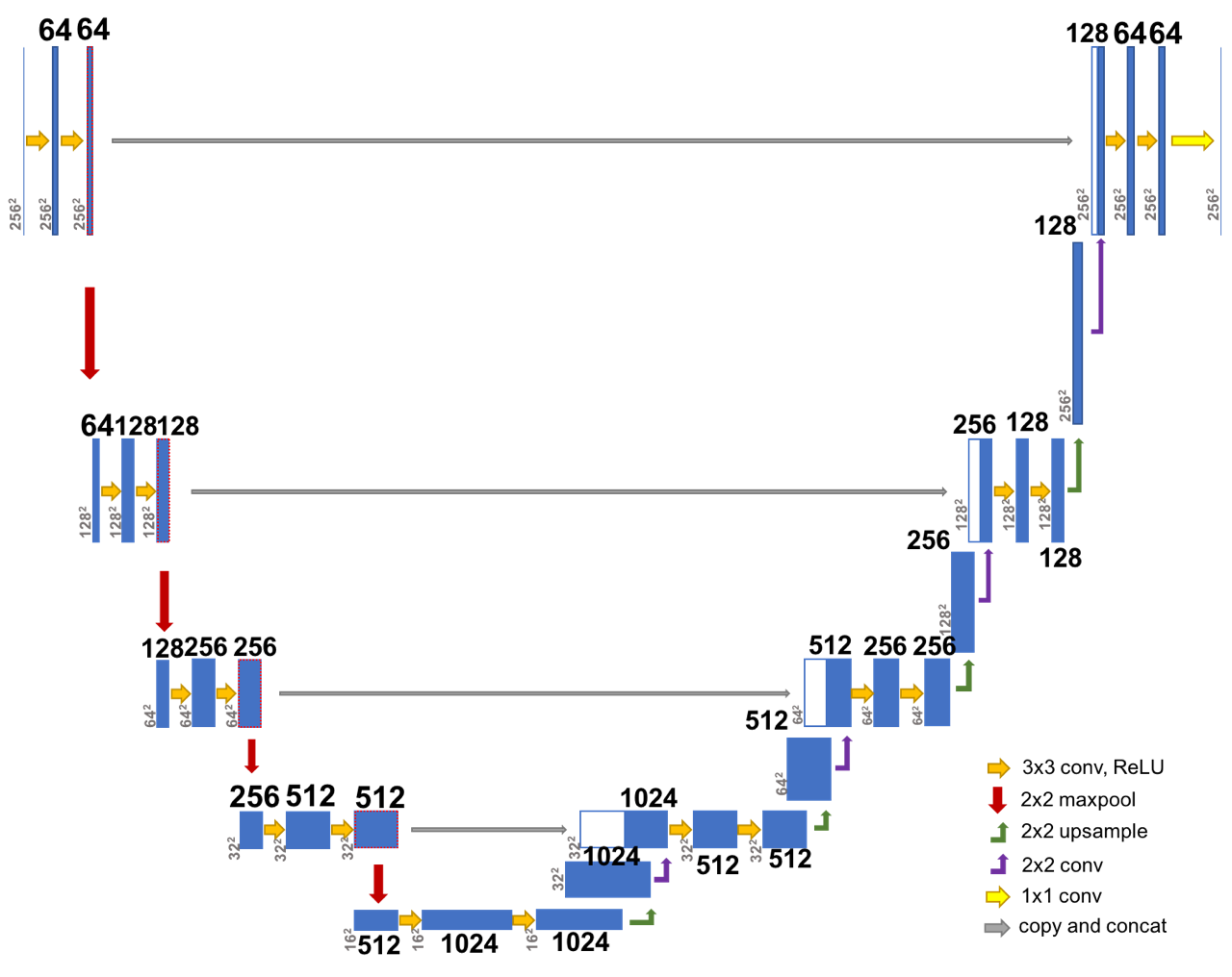
Convolutional neural networks have been applied to a wide variety of computer vision tasks. Recent advances in semantic segmentation have enabled their application to medical image segmentation. While most CNNs use two-dimensional kernels, recent CNN-based publications on medical image segmentation featured three-dimensional kernels, allowing full access to the three-dimensional structure of medical images. Though closely related to semantic segmentation, medical image segmentation includes specific challenges that need to be addressed, such as the scarcity of labelled data, the high class imbalance found in the ground truth and the high memory demand of three-dimensional images. In this work, a CNN-based method with three-dimensional filters is demonstrated and applied to hand and brain MRI. Two modifications to an existing CNN architecture are discussed, along with methods on addressing the aforementioned challenges. While most of the existing literature on medical image segmentation focuses on soft tissue and the major organs, this work is validated on data both from the central nervous system as well as the bones of the hand.
PDF AbstractDatasets
Results from the Paper
 Ranked #2 on
Brain Tumor Segmentation
on BRATS-2015
(using extra training data)


 BraTS 2015
 BraTS 2013