Multi-modal segmentation of 3D brain scans using neural networks
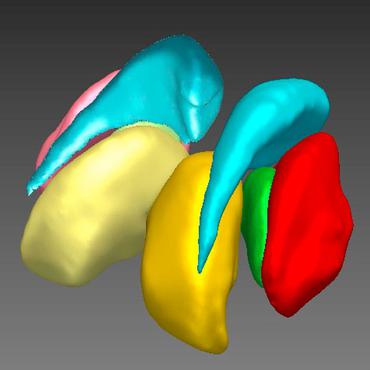
Purpose: To implement a brain segmentation pipeline based on convolutional neural networks, which rapidly segments 3D volumes into 27 anatomical structures. To provide an extensive, comparative study of segmentation performance on various contrasts of magnetic resonance imaging (MRI) and computed tomography (CT) scans. Methods: Deep convolutional neural networks are trained to segment 3D MRI (MPRAGE, DWI, FLAIR) and CT scans. A large database of in total 851 MRI/CT scans is used for neural network training. Training labels are obtained on the MPRAGE contrast and coregistered to the other imaging modalities. The segmentation quality is quantified using the Dice metric for a total of 27 anatomical structures. Dropout sampling is implemented to identify corrupted input scans or low-quality segmentations. Full segmentation of 3D volumes with more than 2 million voxels is obtained in less than 1s of processing time on a graphical processing unit. Results: The best average Dice score is found on $T_1$-weighted MPRAGE ($85.3\pm4.6\,\%$). However, for FLAIR ($80.0\pm7.1\,\%$), DWI ($78.2\pm7.9\,\%$) and CT ($79.1\pm 7.9\,\%$), good-quality segmentation is feasible for most anatomical structures. Corrupted input volumes or low-quality segmentations can be detected using dropout sampling. Conclusion: The flexibility and performance of deep convolutional neural networks enables the direct, real-time segmentation of FLAIR, DWI and CT scans without requiring $T_1$-weighted scans.
PDF Abstract