CNN Based Segmentation of Infarcted Regions in Acute Cerebral Stroke Patients From Computed Tomography Perfusion Imaging
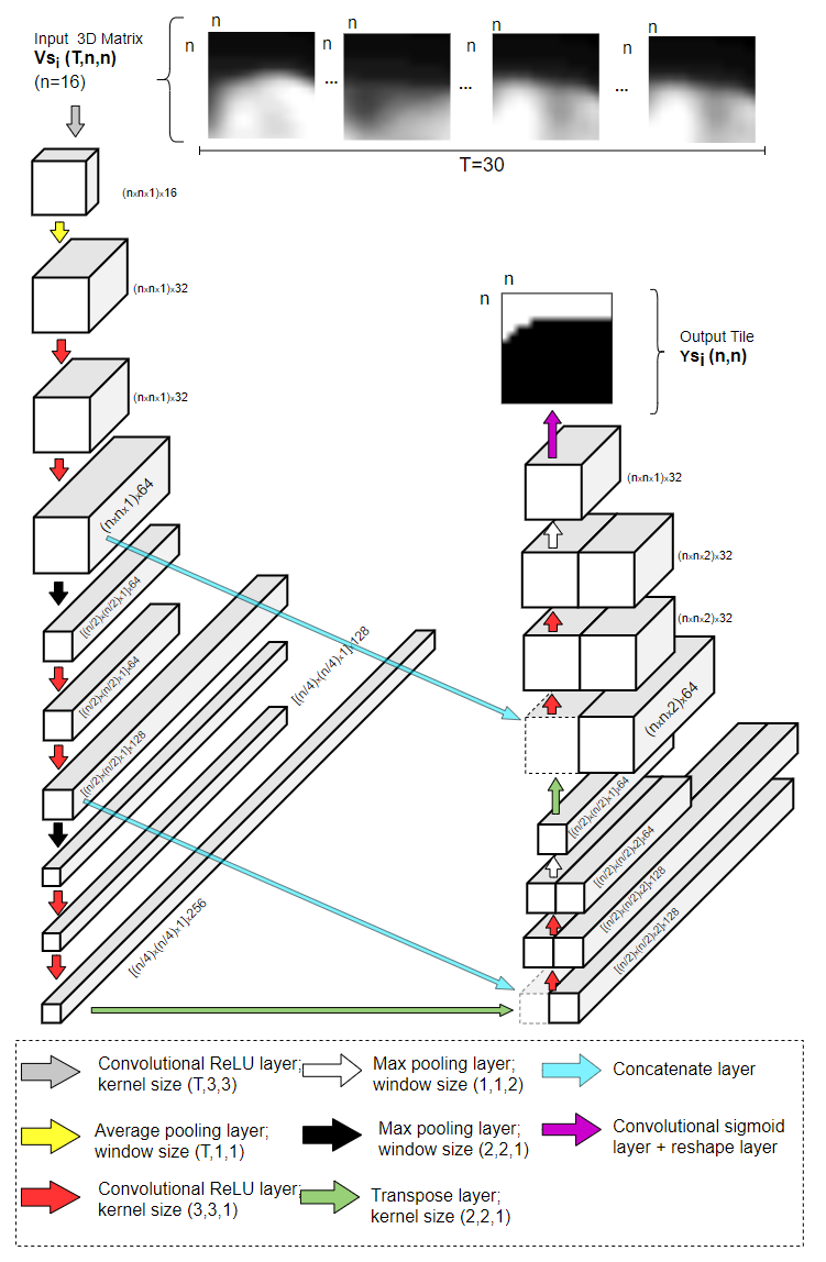
More than 13 million people suffer from ischemic cerebral stroke worldwide each year. Thrombolytic treatment can reduce brain damage but has a narrow treatment window. Computed Tomography Perfusion imaging is a commonly used primary assessment tool for stroke patients, and typically the radiologists will evaluate resulting parametric maps to estimate the affected areas, dead tissue (core), and the surrounding tissue at risk (penumbra), to decide further treatments. Different work has been reported, suggesting thresholds, and semi-automated methods, and in later years deep neural networks, for segmenting infarction areas based on the parametric maps. However, there is no consensus in terms of which thresholds to use, or how to combine the information from the parametric maps, and the presented methods all have limitations in terms of both accuracy and reproducibility. We propose a fully automated convolutional neural network based segmentation method that uses the full four-dimensional computed tomography perfusion dataset as input, rather than the pre-filtered parametric maps. The suggested network is tested on an available dataset as a proof-of-concept, with very encouraging results. Cross-validated results show averaged Dice score of 0.78 and 0.53, and an area under the receiver operating characteristic curve of 0.97 and 0.94 for penumbra and core respectively
PDF Abstract