Deep Learning-based Four-region Lung Segmentation in Chest Radiography for COVID-19 Diagnosis
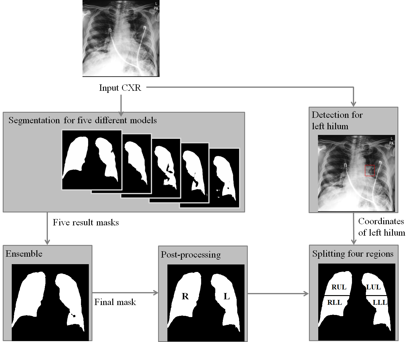
Purpose. Imaging plays an important role in assessing severity of COVID 19 pneumonia. However, semantic interpretation of chest radiography (CXR) findings does not include quantitative description of radiographic opacities. Most current AI assisted CXR image analysis framework do not quantify for regional variations of disease. To address these, we proposed a four region lung segmentation method to assist accurate quantification of COVID 19 pneumonia. Methods. A segmentation model to separate left and right lung is firstly applied, and then a carina and left hilum detection network is used, which are the clinical landmarks to separate the upper and lower lungs. To improve the segmentation performance of COVID 19 images, ensemble strategy incorporating five models is exploited. Using each region, we evaluated the clinical relevance of the proposed method with the Radiographic Assessment of the Quality of Lung Edema (RALE). Results. The proposed ensemble strategy showed dice score of 0.900, which is significantly higher than conventional methods (0.854 0.889). Mean intensities of segmented four regions indicate positive correlation to the extent and density scores of pulmonary opacities under the RALE framework. Conclusion. A deep learning based model in CXR can accurately segment and quantify regional distribution of pulmonary opacities in patients with COVID 19 pneumonia.
PDF Abstract