Multi-template MRI mouse brain atlas (Multi-template MRI mouse brain atlas for both in vivo and ex vivo analysis)

![]()
Mouse Brain MRI atlas (both in-vivo and ex-vivo) (repository relocated from the original webpage)
List of atlases
-
FVB_NCrl: Brain MRI atlas of the wild-type
FVB_NCrlmouse strain (used as the background strain for therTg4510which is a tauopathy model mice express a repressible form of human tau containing the P301L mutation that has been linked with familial frontotemporal dementia.) -
NeAt: Brain MRI atlas of the whld-type
C57BL/6Jmouse strain. Atlas was created based on the originalMRM NeAtmouse brain atlas (template images reoriented and bias-corrected, left/right structure label seperated, and 4th ventricle manual segmentation added). -
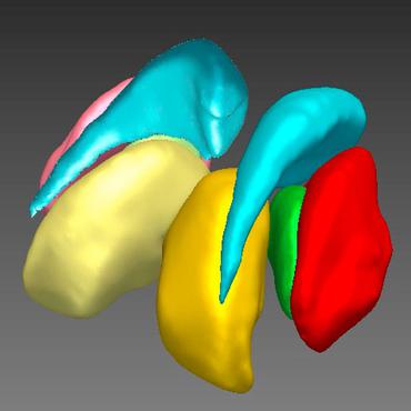
Tc1 Cerebellum: TC1 mouse cerebellar cortical sublayer lobules.This mouse cerebellar atlas can be used for mouse cerebellar morphometry.
Citation
-
If you use the segmented brain structure, or use the atlas along with the automatic mouse brain MRI segmentation tools, we ask you to kindly cite the following papers:
-
Ma D, Cardoso MJ, Modat M, Powell N, Wells J, Holmes H, Wiseman F, Tybulewicz V, Fisher E, Lythgoe MF, Ourselin S. Automatic structural parcellation of mouse brain MRI using multi-atlas label fusion. PloS one. 2014 Jan 27;9(1):e86576. http://journals.plos.org/plosone/article?id=10.1371/journal.pone.0086576
-
Ma D, Holmes HE, Cardoso MJ, Modat M, Harrison IF, Powell NM, O'Callaghan J, Ismail O, Johnson RA, O’Neill MJ, Collins EC, Mirza F. Beg, Karteek Popuri, Mark F. Lythgoe, and Sebastien Ourselin Study the longitudinal in vivo and cross-sectional ex vivo brain volume difference for disease progression and treatment effect on mouse model of tauopathy using automated MRI structural parcellation. Frontiers in Neuroscience. 2019;13:11. https://www.frontiersin.org/articles/10.3389/fnins.2019.00011
-
If you use the brain MR images of the
FVB_NCrlmouse strain (the wildtype background of rTg4510), we ask you to kindly cite the following papers: -
Wells JA, O'Callaghan JM, Holmes HE, Powell NM, Johnson RA, Siow B, Torrealdea F, Ismail O, Walker-Samuel S, Golay X, Rega M. In vivo imaging of tau pathology using multi-parametric quantitative MRI. Neuroimage. 2015 May 1;111:369-78. https://www.sciencedirect.com/science/article/pii/S105381191500124X
-
Holmes HE, Colgan N, Ismail O, Ma D, Powell NM, O'Callaghan JM, Harrison IF, Johnson RA, Murray TK, Ahmed Z, Heggenes M. Imaging the accumulation and suppression of tau pathology using multiparametric MRI. Neurobiology of aging. 2016 Mar 1;39:184-94. https://www.sciencedirect.com/science/article/pii/S0197458015006053
-
Holmes HE, Powell NM, Ma D, Ismail O, Harrison IF, Wells JA, Colgan N, O'Callaghan JM, Johnson RA, Murray TK, Ahmed Z. Comparison of in vivo and ex vivo MRI for the detection of structural abnormalities in a mouse model of tauopathy. Frontiers in neuroinformatics. 2017 Mar 31;11:20. https://www.frontiersin.org/articles/10.3389/fninf.2017.00020/full
-
If you're using the mouse MRI T2* Active Starining Cerebellar atlas, we ask you to please kindly cite the following papers:
- Ma, D., Cardoso, M. J., Zuluaga, M. A., Modat, M., Powell, N. M., Wiseman, F. K., Cleary, J. O., Sinclair, B., Harrison, I. F., Siow, B., Popuri, K., Lee, S., Matsubara, J. A., Sarunic, M. V, Beg, M. F., Tybulewicz, V. L. J., Fisher, E. M. C., Lythgoe, M. F., & Ourselin, S. (2020). Substantially thinner internal granular layer and reduced molecular layer surface in the cerebellum of the Tc1 mouse model of Down Syndrome – a comprehensive morphometric analysis with active staining contrast-enhanced MRI. NeuroImage, 117271. https://doi.org/https://doi.org/10.1016/j.neuroimage.2020.117271
- Ma, D., Cardoso, M. J., Zuluaga, M. A., Modat, M., Powell, N., Wiseman, F., Tybulewicz, V., Fisher, E., Lythgoe, M. F., & Ourselin, S. (2015). Grey Matter Sublayer Thickness Estimation in the Mouse Cerebellum. In Medical Image Computing and Computer Assisted Intervention 2015 (pp. 644–651). https://doi.org/10.1007/978-3-319-24574-4_77
Reference
- For the original information of the
NeAtatlas, please please refer to the website: http://brainatlas.mbi.ufl.edu/, and the following two reference papers: - Ma Yu, Smith David, Hof Patrick R, Foerster Bernd, Hamilton Scott, Blackband Stephen J, Yu Mei, Benveniste Helene In Vivo 3D Digital Atlas Database of the Adult C57BL/6J Mouse Brain by Magnetic Resonance Microscopy. Front. Neuroanat. 2, 1 (2008).
- Ma Yu, Hof P R, Grant S C, Blackband S J, Bennett R, Slatest L, McGuigan M D, Benveniste H A three-dimensional digital atlas database of the adult C57BL/6J mouse brain by magnetic resonance microscopy. Neuroscience 135, 1203–15 (2005).
Funding
The works in this repositories received multiple funding from EPSRC, UCL Leonard Wolfson Experimental Neurology center, Medical Research Council (MRC), the NIHR Biomedical Research Unit (Dementia) at UCL and the National Institute for Health Research University College London Hospitals Biomedical Research center, the UK Regenerative Medicine Platform Safety Hub, and the Kings College London and UCL Comprehensive Cancer Imaging center CRUK & EPSRC in association with the MRC and DoH (England), UCL Faculty of Engineering funding scheme, Alzheimer Society Reseasrch Program from Alzheimer Society Canada, NSERC, CIHR, MSFHR Canada, Eli Lilly and Company, Wellcome Trust, the Francis Crick Institute, Cancer Research UK, and University of Melbourne McKenzie Fellowship.
Papers
| Paper | Code | Results | Date | Stars |
|---|