Medical Object Detection
27 papers with code • 3 benchmarks • 3 datasets
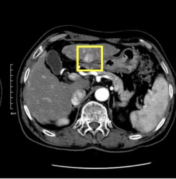
Medical object detection is the task of identifying medical-based objects within an image.
( Image credit: Liver Lesion Detection from Weakly-labeled Multi-phase CT Volumes with a Grouped Single Shot MultiBox Detector )
Most implemented papers
MULAN: Multitask Universal Lesion Analysis Network for Joint Lesion Detection, Tagging, and Segmentation
When reading medical images such as a computed tomography (CT) scan, radiologists generally search across the image to find lesions, characterize and measure them, and then describe them in the radiological report.
Detecting Cancer Metastases on Gigapixel Pathology Images
At 8 false positives per image, we detect 92. 4% of the tumors, relative to 82. 7% by the previous best automated approach.
Retina U-Net: Embarrassingly Simple Exploitation of Segmentation Supervision for Medical Object Detection
The proposed architecture recaptures discarded supervision signals by complementing object detection with an auxiliary task in the form of semantic segmentation without introducing the additional complexity of previously proposed two-stage detectors.
3D Context Enhanced Region-based Convolutional Neural Network for End-to-End Lesion Detection
3D context is known to be helpful in this differentiation task.
Improving RetinaNet for CT Lesion Detection with Dense Masks from Weak RECIST Labels
We propose a highly accurate and efficient one-stage lesion detector, by re-designing a RetinaNet to meet the particular challenges in medical imaging.
Robust End-to-End Focal Liver Lesion Detection using Unregistered Multiphase Computed Tomography Images
The computer-aided diagnosis of focal liver lesions (FLLs) can help improve workflow and enable correct diagnoses; FLL detection is the first step in such a computer-aided diagnosis.
Liver Lesion Detection from Weakly-labeled Multi-phase CT Volumes with a Grouped Single Shot MultiBox Detector
We present a focal liver lesion detection model leveraged by custom-designed multi-phase computed tomography (CT) volumes, which reflects real-world clinical lesion detection practice using a Single Shot MultiBox Detector (SSD).
Attention-Based Deep Neural Networks for Detection of Cancerous and Precancerous Esophagus Tissue on Histopathological Slides
Deep learning-based methods, such as the sliding window approach for cropped-image classification and heuristic aggregation for whole-slide inference, for analyzing histological patterns in high-resolution microscopy images have shown promising results.
MVP-Net: Multi-view FPN with Position-aware Attention for Deep Universal Lesion Detection
In this paper, we propose to incorporate domain knowledge in clinical practice into the model design of universal lesion detectors.
Real-Time Polyp Detection, Localization and Segmentation in Colonoscopy Using Deep Learning
Benchmarking of novel methods can provide a direction to the development of automated polyp detection and segmentation tasks.


 DeepLesion
 SKM-TEA
 RF100
RF100
